Lesion material applied to a glass slide, heat fixed, stained by the method of Gram and examined by light microscopy usually contains D. nodosus cells which have characteristic morphology: large Gram negative bacilli with parallel sides (3-10 μm long and 0.6-0.8 μm wide), often with terminal enlargements (0.8-1.2 μm wide), usually singly but occasionally as pairs joined end to end (Beveridge, 1941). The flora is usually dominated by other species including F. necrophorum, coccobacilli, diptheroids and spirochaetes, some of which may clump around isolated D. nodosus, a phenomenon known as satellitism (Beveridge, 1941).
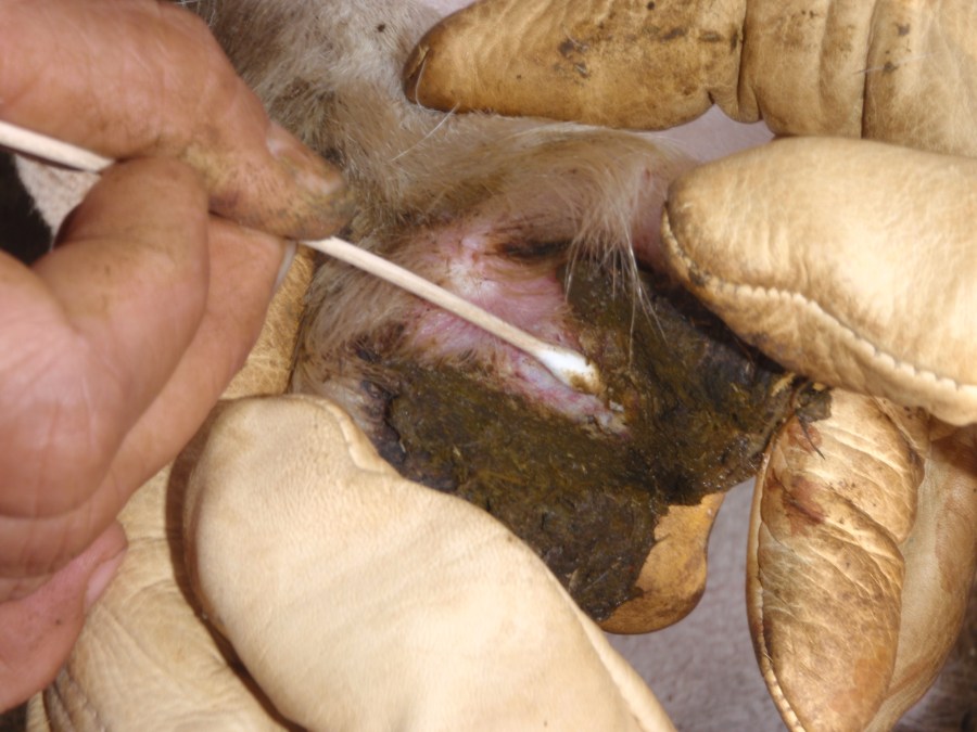
Figure 1. A sample of exudate is taken from the skin-horn junction of an interdigital inflammatory lesion (Score 1 or 2) using a cotton tipped swab. Photo: Richard Whittington

Figure 2. A sample of exudate is taken from the advancing margin of an underrun lesion (Score 3 or 4) using swab stick. The swab can be used to make a smear on a glass slide, inoculate a culture plate or placed into transport medium for later culture, or examined directly using PCR. Photo: John Egerton
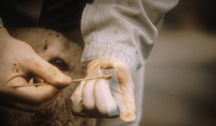
Figure 3. The lesion sample is spread on a glass slide, dried and stained. The same swab can be used to inoculate a culture plate or placed into transport medium for later culture, or examined directly using PCR. Photo: John Egerton
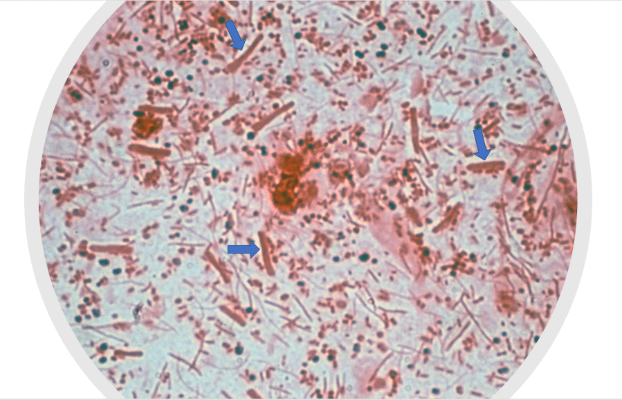
Figure 4. Smear of lesion sample on a glass slide from a case of footrot, stained with Gram. There are a mixture of Gram positive (black) and Gram negative (red) bacteria present. D. nodosus is easily recognised as the largest rod-shaped bacteria in the image (blue arrows). Photo: Richard Whittington
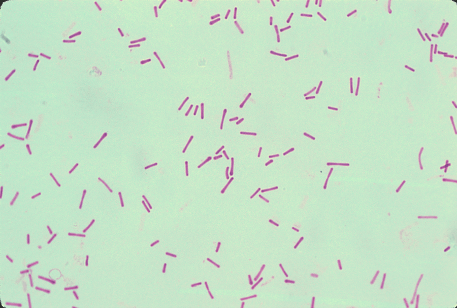
Figure 5. Pure culture of D. nodosus. Gram stain. Photo: Richard Whittington
Go back to: Introduction to lab confirmation
